Can AI Predict Whether a Cancer Cell Will Divide or Die?
A model watched time-lapse videos of cancer cells, including cells given chemotherapy, and learned to guess which would divide and which would die. Where it looked also hints at when the clues appear.
· ~5 min read
The breakdown
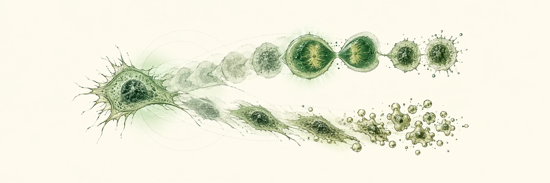
Chemotherapy rarely kills every cancer cell. Cells with identical DNA, in the same dish and at the same dose, can still split into two groups: some die, and others keep dividing and may seed a relapse. Telling which cell will do what, and how early, is a basic question about drug response. A new study trained a deep-learning model to answer it from microscope video alone.1
The gap
Single signals are known to matter. One is p53, a protein that responds to DNA damage. A 2012 study found that cells showing repeated p53 pulses recovered from damage, while cells held in a sustained p53 response frequently entered senescence, a permanent stop in dividing.5
What the authors say was missing is a systematic test of how predictable fate is from several moving signals at once, and when during a cell's life that information shows up. They also say earlier deep-learning work mostly classified cell-cycle stage or modeled tissues, and that Transformer models had not been applied to this chemotherapy setting. We did not independently test that last claim.1
How the model works
The data. The team used a public set of 274 fluorescence time-lapse videos of cancer cells. Frames came every 30 minutes for 93 hours. In 118 videos the cells received cisplatin, a DNA-damaging chemotherapy drug, 48 hours in. Three glowing reporters tracked the cell cycle, a circadian-clock protein, and p53 activity.1, 3 The dataset comes from the same group's earlier cell-tracking preprint.4
Cells were tracked and labeled by hand. A division was marked when a parent cell separated into two daughters, and a death at the first frame showing clear collapse. Table 1 lists 21,898 labeled trajectories: 17,442 ending in division and 4,456 in death. Each cell was cropped around its center from its first appearance up to the frame before the event, so the split or collapse itself was never shown to the model.1
The model. A network called ResNet-50 turns each cropped frame into a list of 2,048 numbers, a compact description of what the image looks like. These lists form a sequence, which goes into a Transformer, a model type built around self-attention. In self-attention, every time point can weigh every other time point, so the model can link a clue early in a video to one near the end.6 A final attention layer gives each frame a weight, which later lets the authors ask which frames mattered.1
This design suits sequences of varying length. A reasonable alternative is hand-measured features such as cell area or shape, and the authors tested a version of that: the same Transformer fed those features instead of image embeddings.1
They split trajectories randomly into 80% training, 10% validation, and 10% test sets. Because dying cells are rarer, they balanced training samples and judged results with balanced accuracy, the average of the share correct within each class, so rare dying cells count as much as common dividing ones.1
What the paper reports
The test set held 2,089 trajectories: 439 deaths and 1,650 divisions. It was drawn at random from the combined labeled dataset. In the full dataset, Table 1 lists 693 untreated controls, 4,722 trajectories from three known cisplatin doses, and 16,483 with no dose information. The paper does not break the test set down by dose, so the scores below are not a treated-only result. The paper restricts its interpretation analyses to cisplatin-treated sequences. Balanced accuracy was 0.94 and the F1 score, another summary of accuracy, was 0.93. The model correctly flagged 401 of 439 deaths (91.3%) and wrongly labeled 3.5% of dividing cells as dying. The hand-feature version scored 0.90 balanced accuracy and 0.877 F1, so images helped, though hand features also did well.1
The authors trimmed frames off the end of each trajectory. With the last 15 frames removed, which is 7.5 hours at 30-minute spacing, balanced accuracy was still 0.903. At 20 or more frames removed it fell steeply, reaching 0.565 at 40. Using only the final frame gave 0.713.1
The two fates behaved differently. Division could be recognized from very few late frames: recall, the share of true divisions caught, reached 0.918 using the last four frames. Death needed far more context, reaching a recall of 0.907 only with about 80 frames.1
To check that attention reflects real reliance, the authors masked frames. Hiding half the frames with the highest weights cut balanced accuracy to 0.836. Hiding random frames left 0.912, and hiding the lowest-weight frames left 0.931.1
The weights themselves told a similar story. For dividing cells, attention piled up at the end: normalized weight was 0.62 at the second-to-last frame and 0.22 ten frames earlier. For dying cells it was spread out: 0.88 at the last frame, about 0.42 thirty frames earlier, and 0.07 at 45 frames.1
Comparing high- and low-attention frames, p53 intensity showed the largest effect for both fates, measured with Cliff's delta, a statistic of how often values in one group exceed the other. Frames before division showed larger, rounder cells, while frames before death showed larger cells that did not round. The cell-cycle reporter showed no significant difference.1
Caveats. The paper describes a random split of trajectories and does not say whether cells from one video were kept together. Cells in a video share a dish and imaging conditions, so performance on a new experiment is unknown. This is our reading, not a flaw the authors report. The untreated control had only 41 deaths, so the authors did not interpret those estimates.1
Attention is also a debated explanation. Work on language models found that attention weights often do not line up with other importance measures.7 The masking test supports the authors' use here, but it shows what the model relies on, not what the cell is doing. The authors suggest testing features by removing them from a model.1
Future directions
These are proposals anchored to the paper's limits. As of October 19, 2026, each idea was searched in PubMed, bioRxiv, arXiv, and the web, then given a separate adversarial check. Anyone picking up one of these ideas should repeat a fresh literature search before committing to a project.
1. Test on whole held-out experiments. Scoring the model on complete videos, a different dose, or a different drug or cell line would show whether it generalizes. The searches did not surface such a study for a division-versus-death classifier. Related work probes cell-fate predictors by virtually changing their inputs.8
2. Compare attention with other explanation methods. The authors validate attention by masking frames. Gradient-based attribution could be run on the same model and compared. The searches did not surface such a comparison for time-lapse cell-fate models. The closest adjacent work is the virtual perturbation study.8
3. Link predicted fate to gene activity. The authors propose profiling cells with single-cell transcriptomics at the time points when fate is decided. This is partly explored. A 2025 study predicted survival or death after heat stress from images, then picked cells for single-cell RNA sequencing and compared predicted-dead and predicted-survivor cells.9 The searches did not surface this for division versus death after chemotherapy.
Why it matters
Time-lapse imaging is usually descriptive. This work shows it can also be predictive, and a model's focus can suggest when fate takes shape. The open question is whether it holds on new experiments.
References & paper details
Paper details
- Venue: PLOS Computational Biology 22(10): e1014813.1
- Publication date: October 12, 2026, per PubMed. An arXiv preprint of the same title was posted March 17, 2026.2
- Version analyzed: The journal version's full text via PubMed Central.
- Peer review: Peer reviewed.
- Study type: Computational method development, using a public imaging dataset.
- Data and code: The arXiv version lists code at github.com/bozeklab/Cell-Fate-Prediction and the raw dataset at doi.org/10.4126/FRL01-006526608.2
- License: PubMed lists the article as open access under a Creative Commons Attribution license. This post is an independent summary.
References
1. Central research paper
Florian Bürger, Martim Dias Gomes, Adrián E. Granada, Noémie Moreau, and Katarzyna Bozek. Understanding cell fate decisions with temporal attention. PLOS Computational Biology 22(10): e1014813, 2026. DOI: 10.1371/journal.pcbi.1014813.
2. arXiv preprint
Bürger and colleagues. Understanding Cell Fate Decisions with Temporal Attention. arXiv:2603.16562, March 2026. Preprint version, not peer reviewed. arxiv.org/abs/2603.16562.
3. Code and dataset
Cell-Fate-Prediction repository and raw dataset, as listed in the preprint.
4. TransientTrack
Florian Bürger, Martim Dias Gomes, Nica Gutu, Adrián E. Granada, Noémie Moreau, and Katarzyna Bozek. TransientTrack: Advanced Multi-Object Tracking and Classification of Cancer Cells with Transient Fluorescent Signals. arXiv:2512.01885. Preprint, not peer reviewed. arxiv.org/abs/2512.01885.
5. p53 dynamics
Jeremy E. Purvis, Kyle W. Karhohs, Caroline Mock, Eric Batchelor, Alexander Loewer, and Galit Lahav. p53 dynamics control cell fate. Science 336(6087): 1440-1444, 2012. DOI: 10.1126/science.1218351.
6. The Transformer
Ashish Vaswani and colleagues. Attention Is All You Need. arXiv:1706.03762, 2017. arxiv.org/abs/1706.03762.
7. Attention is not Explanation
Sarthak Jain and Byron C. Wallace. Attention is not Explanation. arXiv:1902.10186, 2019. arxiv.org/abs/1902.10186.
8. Virtual perturbations
Christopher J. Soelistyo, Guillaume Charras, and Alan R. Lowe. Virtual Perturbations to Assess Explainability of Deep-Learning Based Cell Fate Predictors. IEEE/CVF International Conference on Computer Vision Workshops, 2023, pp. 3973-3982. mlanthology.org entry.
9. Fate prediction plus RNA sequencing
Tomoaki Okaniwa, Kirill Kryukov, and Katsuyuki Shiroguchi. Finding differentially expressed genes between cell fates predicted by image-based deep learning. Biophysics and Physicobiology 22(4): e220022, 2025. DOI: 10.2142/biophysico.bppb-v22.0022.
Get the next breakdown
Subscribe for one email per issue — methods, limits, and what it actually means.
Related breakdowns
October 2026
Modeling Lab Bias Sharpens the Search for Overlooked Reading Frames
Cells read short stretches of genetic code that gene lists leave out, and the lab method used to find them can smear its own signal. A new tool, RiboBA, models that smearing instead of ignoring it.
Read breakdown →
October 2026
AI Beat Docking at Poses, but Not Consistently at Finding Hits
A UCSF team tested three AI structure predictors on hundreds of brand-new crystal structures, then on long lists of candidate drugs. The AI won at drawing poses but did not consistently improve hit ranking.
Read breakdown →
October 2026
How Scientists Sorted 2.8 Million Bacterial Gene Clusters in Under 18 Hours
Bacteria keep related genes together in groups, and scientists now have millions of them to compare. IGUA groups them quickly by removing duplicates first, then comparing what is left.
Read breakdown →